Clinical Neuroanatomy (Yilmazer-Hanke)
Group members:
Prof. Dr. D. Yilmazer-Hanke (Head / Leitung), Dr. Elisa Tuzzi (Postdoctoral Researcher), G. Ehmke (MTA), S. Feldengut (MTA), P. Häring (MTA)
The focus of the Clinical Anatomy Section is (I) to apply modern techniques in human brain research, (II) to perform clinicopathological correlations with clinical symptoms, biomarkers and imaging in conjunction with experimental translational studies, and (III) to educate young scientists and professionals, who work in various medical disciplines and neuroanatomical research.
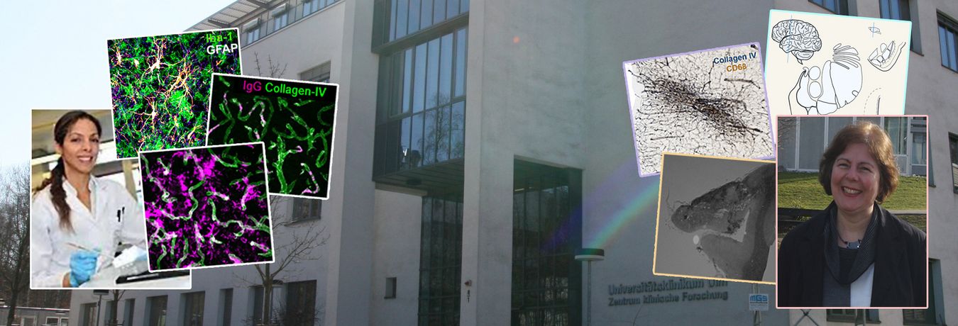
Research areas of interest include human neurodegenerative diseases, cerebral microangiopathies and basic research in structural, functional and molecular neuroanatomy. Examples of our research can be found in the display of the Tissue Archive.
The Clinical Neuroanatomy further coordinates autopsies for patients in the Neurology Department including Neurogeriatrics and Neurological Rehabilitation.
We offer external teaching courses to graduate students and trainees in neuropathology supported by professional societies and associations. Teaching areas at University level include thesis supervision and courses, lectures and laboratory practicals in the Masters in Molecular and Translational Neuroscience (MTN) and Masters in Molecular Medicine (MMU) Programs.
Research
The major focus are cerebral microangiopathies and the link to neurodegeneration in the human brain. These diseases represent two extremes of a spectrum of cerebral pathologies that cause cognitive decline and dementia in the aging population. Mechanisms leading to these diseases and the impact of aging on their development are examined in experimental models. Further investigations include the study of the functional, molecular, genetic and pathological anatomy of the brain with an emphasis on prefrontal-amygdala-hippocampal networks and downstream brain areas such as the hypothalamus and the brainstem.
Microangiopathies cause microinfarctions, microbleeds and a variety of other lesions in the brain. In the clinical setting, cortical cerebral microinfarcts often escape detection in patients, owing to the comparatively low magnetic field strengths and resolution of clinical scanners that are in use for in vivo imaging. Moreover, due to the large size of the human brain a large proportion of microinfarcts remains undetected in routine neuropathological investigations. These observations suggest that the prevalence of microinfarcts may be higher than expected. Therefore, we are working on developing new histological tools and in vivo / ex vivo imaging procedures that allow a better characterization and detection of cortical cerebral microinfarcts and other vascular pathologies in the brain.
Another cerebral vascular focus encompasses investigations on white matter hyperintensities (WMH), a common finding in human brain imaging. The pathological correlate of WMH are white matter lesions (WML) in the brain. In the human brain, we recently were able to identify and characterize vascular bags that are filled with plasma proteins and various stages of string vessels in WML. This indicates breakdown of the blood-brain-barrier (BBB) with extravasation of plasma proteins to vascular bags.
The breakdown of the BBB, and also of the blood-spinal cord-barrier (BSCB), is a common finding in many neurodegenerative diseases including Alzheimer’s disease, Parkinson’s disease and amyotrophic lateral sclerosis (ALS). The disruption of the BBB and BSCB lead to neuronal dysfunction and glial activation and cause tissue damage in the white matter associated with demyelination and axonopathy, probably leading to the development of WML. The aim of ongoing studies is therefore to determine the prevalence and mechanisms that cause the development of various vascular pathologies in the human brain. With the aid of experimental models that employ genetic and chemogenetic tools, the effects of BBB and BSCB disruption are studied in different cell populations.
The research strategy is based on the analysis of limbic brain regions and related areas relevant for the formation of emotional memory in neurodegenerative diseases, neuropsychiatric conditions such anxiety-related or stress-related disorders and temporal lobe epilepsy. Experiments are performed in the human brain and in genetically defined rodent strains. Studies at molecular, cellular and systems level are aimed at developing new therapeutic strategies including the application of peptides and drugs.
Our long term goal remains the identification of novel biomarkers and targets for the diagnostics and therapy of cerebral microangiopathies, which is critical for a successful treatment of patients with dementia. This requires clinical monitoring of patients with microangiopathies, investigations on cerebrospinal fluid biomarkers and the advancement of imaging techniques. Therefore, our group works in close collaboration with the clinical cerebrovascular, clinical neuropsychology, neuroimaging, neurochemistry and metabolomics units in the Neurology Department. Mechanistic experimental studies are performed in collaboration with various groups at the University and the DZNE. External collaborations include studies on diseases that affect the limbic system and amygdala, novel imaging methods, clinical cerebrovascular research and neuropathology.
Baustein 3.2. Principal Investigator: Ouali Alami N. "Experimental mouse models of human cerebral small vessel disease" Funding period 2020/21, 50.000,- €
- NEBRASKA/ LB595 -Cancer and Smoking Disease Research Program Development. Principal Investigator: Yilmazer-Hanke D. “Regulation of ITIH3 by nicotine and tobacco smoke through the CD44 receptor”. 2015 – 2017 $ 120,000 (completed on Oct 31, 2016 as non-transferable to Ulm University)
- NIH-NIGMS (CoBRE) “Molecular Biology of Neurosensory Systems” 8P20GM103471-09 (Subaward No.: 34-5507-2020-109 to Yilmazer-Hanke D.), 5P20GM103471-10 (Subaward No.: 34-5507-2020-010 to Yilmazer-Hanke D.) and several Vouchers for usage of Core Facilities. “Cis-acting expression quantitative trait loci (eQTLs) in recombinant inbred lines differing in fear: A microarray study”. 2012-2014 $ 136,597
- NEBRASKA/LB692 Funds. Principal Investigator: Yilmazer-Hanke D. “Role of Limbic Areas in Emotional Changes and in Epilepsy”. 2011-2014 $ 300,000
Link zu Pubmed -> Link
- Yilmazer-Hanke D, Mayer T, Müller HP, Neugebauer H, Abaei A, Scheuerle A, Weis J, Forsberg KME, Althaus K, Meier J, Ludolph AC, Del Tredici K, Braak H, Kassubek J, Rasche V. Histological correlates of postmortem ultra-high-resolution single-section MRI in cortical cerebral microinfarcts. Acta Neuropathol Commun, 8(1):33 (2020)
- Ouali Alami N, Tang L, Wiesner D, Commisso B, Bayer D, Weishaupt JH, Dupuis L, Wong PC, Baumann B, Wirth T, Boeckers TM, Yilmazer-Hanke D, Ludolph AC, Roselli (2020). Multiplexed chemogenetics in astrocytes and motoneurons restore Blood-Spinal Cord-Barrier in ALS. Life Sci Alliance. 3(11):e201900571 (2020).
- Abdelhak A, Huss A, Brueck A, Sebert U, Mayer B, Tumani H, Otto M, Yilmazer-Hanke D, Ludolph AC, Kassubek J, Pinkhardt E, Neugebauer H. Optical coherence tomography-based assessment of retinal vascular pathology in cerebral small vessel disease. doi.org/10.1186/s42466-020-00062-4 Neurol Res Pract. 2:13 (2020).
- Lutz AK, Pfaender S, Incearap B, Ioannidis V, Ottonelli I, Föhr KJ, Cammerer J, Zoller M, Higelin J, Giona F, Stetter M, Stoecker N, Ouali Alami N, Schön M, Orth M, Liebau S, Barbi G, Grabrucker AM, Delorme R, Fauler M, Mayer B, Jesse S, Roselli F, Ludolph AC, Bourgeron T, Verpelli C, Demestre M, Boeckers TM. Autism-associated SHANK3 mutations impair maturation of neuromuscular junctions and striated muscles. Sci Transl Med. 12(547), eaaz3267 (2020).
- Bączyk M, Ouali Alami N, Delestrée N, Martinot C, Tang L, Commisso B, Bayer D, Doisne N, Frankel W, Manuel M, Roselli F, Zytnicki D. Synaptic restoration by cAMP/PKA drives activity-dependent neuroprotection to motoneurons in ALS. J Exp Med. 217(8):e20191734 (2020)
- Bruno C, Sieverding K, Freischmidt A, Satoh T, Walther P, Mayer B, Ludolph AC, Akira S, Yilmazer-Hanke D, Danzer KM, Lobsiger CS, Brenner D, Weishaupt JH. Haploinsufficiency of TBK1 prepones age-associated neuroinflammatory changes without causing motor neuron degeneration in aged mice. Brain Commun. (2020) Epub 2020 Aug 21. DOI: 10.1093/braincomms/fcaa133
- Wickramasekara RN, Bockman C, Hanke J, Schwegler H, McGee J, Walsh E, Yilmazer-Hanke D. Alpha2-adrenergic dysregulation in congenic DxH recombinant inbred mice selectively bred for a high fear-sensitized (H-FSS) startle response. Pharmacol Biochem Behav 188:172835 (2020)
- Datzmann T, Kapapa T, Scheuerle A, McCook O., Merz T, Unmuth S, Hoffmann A, Mathieu R, Mayer S, Mauer UM, Röhrer S, Yilmazer-Hanke D, Möller P, Nussbaum BL, Calzia E, Gröger M, Hartmann C, Radermacher P, Wepler M. In-depth characterization of a long-term, resuscitated model of acute subdural hematoma-induced brain injury. J Neurosurg, 2019 Dec 20:1-12.
- Stefanits H, Milenkovic I, Mahr N, Pataraia E, Baumgartner C, Hainfellner JA, Kovacs GG, Kasprian G, Sieghart W, Yilmazer-Hanke D*, Czech T* (* Joint senior and corresponding author). Alterations in GABAA receptor subunit expression in the amygdala and entorhinal cortex in human temporal lobe epilepsy. J Neuropathol Exp Neurol 78:1022-1048 (2019)
- Oeckl P, Weydt P, Steinacker P, Anderl-Straub S, Nordin F, Volk AE, Diehl-Schmid J, Andersen P, Kornhuber J, Danek A, Fassbender K, Fliessbach K, German Consortium for Frontotemporal Lobar Degeneration, Jahn H, Lauer M, Müller K, Knehr A, Prudlo J, Schneider A, Thal DR, Yilmazer-Hanke D, Weishaupt JH, Ludolph AC, Otto M. Different neuroinflammatory profile in amyotrophic lateral sclerosis and frontotemporal dementia is linked to the clinical phase. J. Neurol Neurosurg Psychiatry 90:4-10 (2019).
- Ouali Alami N, Schurr C, Olde Heuvel F, Tang L, Li Q, Tasdogan A, Kimbara A, Nettekoven M, Ottaviani G, Raposo C, Röver S, Rogers-Evans M, Rothenhäusler B, Ullmer C, Fingerle J, Grether U, Knuesel I, Boeckers TM, Ludolph A, Wirth T, Roselli F, Baumann B. NF-κB activation in astrocytes drives a stage-specific beneficial neuroimmunological response in ALS. EMBO J 37(16). pii: e98697 (2018).
- Braak H, Feldengut S, Kassubek J, Yilmazer-Hanke D, Del Tredici K. Two histological methods for recognition and study of cortical microinfarcts in thick sections. Eur J Histochem Dec 20;62(4) (2018).
- Forsberg KME, Zhang Y, Reiners J, Ander M, Niedermayer A, Neugebauer H, Weiss J, Katona I, Ludolph AC, Del Tredici K, Braak H, Yilmazer-Hanke D. Endothelial damage, vascular bagging and remodeling of the microvascular bed in human deep white matter lesions. Acta Neuropathol Commun Nov 23;6(1):128 (2018).
- Wickramasekara RN, Morrill S, Farhat Y, Smith SJ, Yilmazer-Hanke D. Glutathione and inter-α-trypsin inhibitor heavy chain 3 (Itih3) mRNA levels in nicotine-treated Cd44 knockout mice. Toxicol Rep 5:759–764 (2018).
- Steinacker P, Verde F, Fang L, Feneberg E, Oeckl P, Roeber S, Anderl-Straub S, Danek A, Diehl-Schmid J, Fassbender K, Fliessbach K, Foerstl H, Giese A, Jahn H, Kassubek J, Kornhuber J, Landwehrmeyer GB, Lauer M, Pinkhardt EH, Prudlo J, Rosenbohm A, Schneider A, Schroeter ML, Tumani H, von Arnim CAF, Weishaupt J, Weydt P, Ludolph AC, Yilmazer Hanke D, Otto M; FTLDc study group. Chitotriosidase (CHIT1) is increased in microglia and macrophages in spinal cord of amyotrophic lateral sclerosis and cerebrospinal fluid levels correlate with disease severity and progression. J Neurol Neurosurg Psychiatry 89:239-47 (2018).
- Stefanits H, Milenkovic I, Mahr N, Pataraia E, Hainfellner JA, Kovacs GG, Sieghart W, Yilmazer-Hanke D*, Czech T* (* Joint senior and corresponding author). GABA(A) receptor subunits in the human amygdala and hippocampus: Immunohistochemical distribution of 7 subunits. J Comp Neurol 526:324-48 (2018).
- O'Loughlin E, Pakan JMP, Yilmazer-Hanke D, McDermott KW. Acute in utero exposure to lipopolysaccharide induces inflammation in the pre- and postnatal brain and alters the glial cytoarchitecture in the developing amygdala. J Neuroinflammation 14:212 (2017)
- Yilmazer-Hanke D, Eliava M, Hanke J, Schwegler H, Asan E. Density of acetylcholine esterase (AchE) and tyrosine hydroxylase (TH) containing fibers in the amygdala of roman high- and low-avoidance rats. Neurosci Lett 632, 114-8 (2016)
- Yilmazer-Hanke D, O’Loughlin E, McDermott K. Contribution of amygdala pathology to comormid emotional disturbances in temporal lobe epilepsy. J Neurosci Res. 94:486-503 (2016)
- Sommariva E, Brambilla S, Carbucicchio C, Gambini E, Meraviglia V, Dello Russo A, Farina FM, Casella M, Catto V, Pontone G, Chiesa M, Stadiotti I, Cogliati E, Paolin A, Ouali Alami N, Preziuso C, d'Amati G, Colombo GI, Rossini A, Capogrossi MC, … Pompilio G. Cardiac mesenchymal stromal cells are a source of adipocytes in arrhythmogenic cardiomyopathy. Eur Heart J. 37:1835–1846 (2016)
- Amygdala. Yilmazer-Hanke D. In: Arthur W. Toga (Editor). Brain Mapping: An Encyclopedic Reference, Vol. 2, pp. 341-346. Academic Press: Elsevier, London, ISBN 978-0-12-397025-1 (2015).
- Insights into the Amygdala: Structure, Function and Implications for Disorders. D. Yilmazer-Hanke (Editor). Hauppauge, NY, US: Nova Science Publishers (2012).
- Connections of the rodent central nucleus of amygdala: A functional view. Yilmazer-Hanke, D., Fritz, R., D’Hanis, W., Schwegler, H., Linke, R. In: D. Yilmazer-Hanke (Ed). Insights into the Amygdala: Structure, Function and Implications for Disorders. Hauppauge, NY, US: Nova Science Publishers, pp. 93-138 (2012).
- Amygdala (Chapter 22). Yilmazer-Hanke D. In: Paxinos G, Mai J.K. (eds) The Human Nervous System, 3rd Edition. Academic Press (Elsevier Ltd), London, pp.759-834 (2012).
- Effects of embryo transfer on the phenotype. Rose C., Hanke J., Schwegler H., Yilmazer-Hanke D. Advances in Medicine and Biology, Vol. 39, Nova Science Publishers Inc., pp. 1-47 (2011)
- Schwegler H., Yilmazer-Hanke D., Roskoden T., Crusio W.E. Die Wirkung transienter postnataler Hyperthyreose auf die Entwicklung, Morphologie und Funktion von limbischen Strukturen bei Ratte und Maus. In: Lipinski Ch.G., Braus D.F. (eds) Hippocampus. Klinisch relevante Schlüsselfunktionen. Hippocampus Verlag, Bad Honnef, pp. 39-55 (2004).
- de Vos R.A.I., Jansen E.N.H., Yilmazer D., Braak H., Braak E. Pathological and clinical features of Parkinson`s disease with and without dementia. In: Perry, R.H., McKeith, I.G., Perry, E.K. (eds) Dementia with Lewy bodies. Clinical, pathological, and treatment issues. Cambridge University Press, Cambridge, New York, Melbourne, pp. 255-267 (1996).
The Clinical Neuroanatomy coordinates autopsies for patients in the Neurology Department including Neurogeriatrics and Neurological Rehabilitation.
At present, we are actively recruiting patients for autopsies with neurovascular diseases, cerebral microangiopathies and vascular dementia. Autopsies from all familial and sporadic neurological diseases including neurodegenerative diseases such as Amyotrophic Lateral Sclerosis (ALS), Alzheimer’s disease, frontotemporal Dementia (FTD), Parkinson’s disease and other movement disorders are accepted.
The ultimate diagnosis and underlying cause of cerebral microangiopathies, dementias and other neurodegenerative diseases can only be determined in postmortem examination. The goal of diagnostic investigations and research is to perform clinicopathological correlations to develop new treatment strategies for patients.
Patients, who are interested in donating tissues and organs to the Brain bank at Ulm University, can contact us under hirnbank@uni-ulm.de.
Die Klinische Neuroanatomie koordiniert Autopsien für Patienten in der Neurologie, Neurogeriatrie und Neurologischer Rehabilitation.
Zurzeit rekrutieren wir Patienten für Autopsien mit neurovaskulären Erkrankungen, zerebralen Mikroangiopathien und mit vaskulärer Demenz. Auch werden Autopsien von Patienten mit anderen familiären oder sporadischen neurologischen und neurodegenerativen Erkrankungen wie Amyotrophe Lateral Sklerose (ALS), M. Alzheimer, frontotemporal Demenz (FTD), M. Parkinson und anderen Bewegungsstörungen angenommen.
Die endgültige Diagnose von zerebralen Mikroangiopathien, Demenzen und neurodegenerativen Erkrankungen kann nur durch eine postmortale Untersuchung gestellt werden. Das Ziel der postmortalen Diagnostik und von wissenschaftlichen Untersuchungen ist die Durchführung klinisch-pathologischer Korrelationsstudien, damit neue Behandlungsstrategien entwickelt werden können.
Patienten, die Interesse an einer postmortalen Gewebe oder Organspende an die Hirnbank der Universität Ulm haben, können uns unter der Emailadresse hirnbank@uni-ulm.de kontaktieren.
Since 2018 the Clinical Neuroanatomy offers external teaching courses on the anatomy and pathology of the human nervous system, which are coordinated and supported by the German Neuroscience Society, the Neurowissenschaftliche Gesellschaft (NWG).
In the Translational Neuroanatomy and Pathology course, students are introduced to pathological changes and the staging of human neurodegenerative diseases in lectures and in histology sessions. These findings are then compared to pathological alterations in current state-of-the-art research models. This course is geared towards training graduate students, medical students and undergraduate students with a background in human neurobiology or related discipline. In hands-on laboratory practicals, students also acquire skills that allow them to get insights to laboratory procedures used in human brain research including Braak’s techniques.
The Pathoanatomy of the Human Nervous System course is complementary to the translational course. It provides a deeper knowledge of pathological changes in the human brain also suitable for medical doctors, who are Trainees in Neuropathology. In lectures, histology sessions and laboratory practicals, students and colleagues have the opportunity to learn about advanced research techniques for studying the human brain, to learn about pathologically relevant neuroanatomical concepts, and to perform scorings and Braak’s staging in human neurodegenerative diseases.
https://nwg-info.de/aktivitaeten/kurse_workshops/2020
At University level, the Clinical Neuroanatomy contributes to teaching in the Molecular and Translational Neuroscience (MTN) and Masters in Molecular Medicine (MMU) Programs. Courses taught in MTN include the coordination and delivery of the lecture series "MEDex30: Introduction to Human Neuroanatomy", and lectures on microangiopathies in "MEDex19: Neurological Diseases", and on histological Techniques to image the brain in "MEDex14: Brain Imaging and Neuroanatomy". The MTN students further receive training in human brain research during short (1-wk.) and long (4-wks.) laboratory practicals. In the MMU program, graduate students take 4-wk. laboratory practicals in the course “Signaling Pathways in Stem Cells, Development and Aging”.
The Clinical Neuroanatomy also offers ample opportunities for laboratory research to a variety of students. Bachelor in Science (B.Sc.) and Master in Science (M.Sc.) students in the MTN and MMU programs have successfully graduated upon completion of their laboratory work and thesis in Clinical Neuroanatomy. Numerous engaged medical and dental students also work in Clinical Neuroanatomy toward completion of their doctoral dissertation.
Students, who are interested in a B.Sc. or M.Sc. thesis or doctoral dissertation, please contact us under:
Prof. Dr. Deniz Yilmazer-Hanke
Clinical Neuroanatomy, Neurology
University of Ulm
Tel: +49 (0)731 500-63157 /-63158
Email: deniz.yilmazer-hanke@uni-ulm.de
Contact / Kontakt
Telefon +49 (0)731 500-63157
Prof. Dr. Deniz Yilmazer-Hanke
Clinical Neuroanatomy, Neurology
University of Ulm
Helmholtzstrasse 8/1
89081 Ulm
Team:
Dr. Elisa Tuzzi (Postdoc)
Gabriele Ehmke (MTA)
Simone Feldengut (MTA)
Patricia Häring (MTA)