Preclinical research
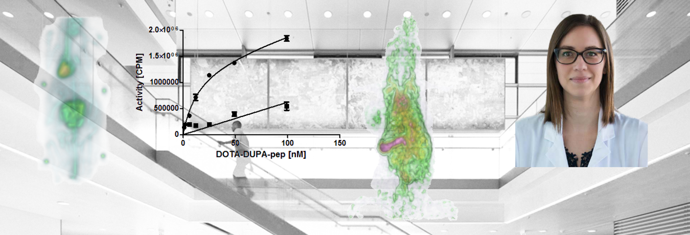
Main research areas in the field of nuclear medicine include the development, evaluation and optimization of oncologic tracers, e.g. for the diagnosis and therapy of prostate carcinoma or leukemias. PSMA-specific peptides are among the main areas of research and are successfully analyzed using established methods.
The diagnosis and analysis of neurodegenerative diseases using novel tau-specific peptides is also the focus of research in the Department of Nuclear Medicine. The accumulation and specificity are mainly studied in vitro and in tissue sections.
In the field of preclinical research, state-of-the-art methods are used in the Department of Nuclear Medicine to successfully work on complex interdisciplinary projects. A broad spectrum of cell lines and great expertise in cultivation and maintenance form the basis for in vitro studies. These include, for example, specific binding studies or time-dependent analyses of the uptake of radiolabeled peptides, antibodies and other compounds. Modern technology, e.g. a cutting-edge chromatography system for protein purification or a flow cytometer for cell studies with fluorescent samples are available in the department.
A digital autoradiography system with a resolution of up to 50 µm is available for autoradiographic analyses, e.g. in histological specimens, protein gels or for the verification of radiolabeling. In addition to the excellent resolution of this imaging technique, quantifiability is also an important advantage.

In the Department of Nuclear Medicine there is a high interest in alternative methods to animal experiments. A technique that is already in use and will be further established are in ovo studies on the HET-CAM (Hen's egg test - chorioallantoic membrane) model. Therefor tumor xenografts are established on the membrane in the hen's egg and biodistribution as well as tumor accumulation studies can be performed to analyze the specificity of substances independently of animal experiments.
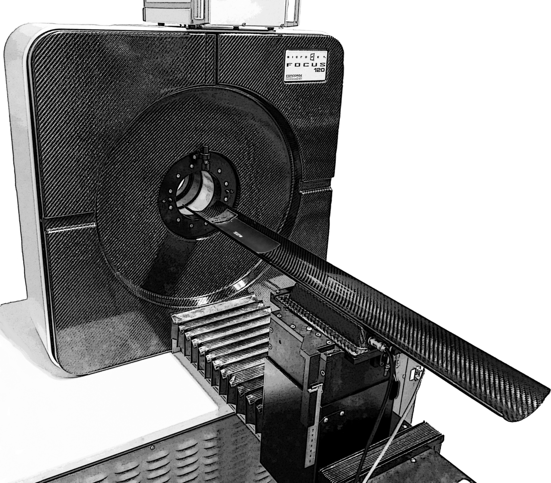
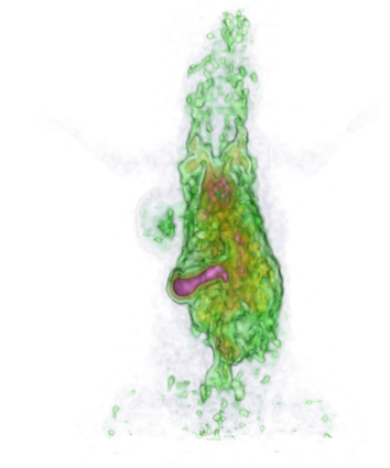
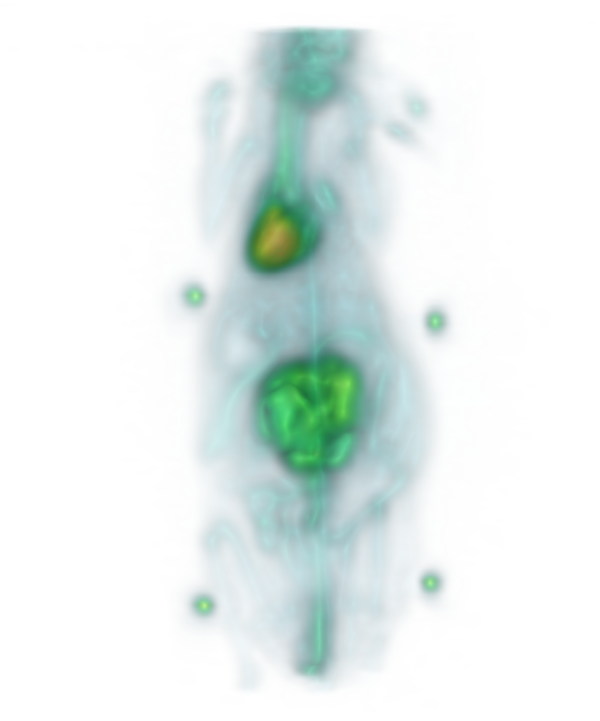
A small animal PET scanner (Focus120) from Siemens is used for the studies of biodistribution, accumulation and efficacy of radiolabeled samples. In close collaboration with the Core Facility for Small Animal Imaging, multi-modality imaging is realized with high-resolution anatomical MR data acquired with a Bruker 11.7 T scanner. The superposition of the image data of both modalities allows an excellent anatomical localization of the signals of the highly sensitive PET scans for the detection of trace amounts of pharmacological substances.
In cooperation with the Institute of Inorganic Chemistry 2 of Prof. Mika Lindén, research is focused on the optimization and modification of nanoparticles, in particular mesoporous silica nanoparticles, for application in imaging and therapy, especially in oncology. New syntheses, variable surface modifications and improved biodistribution combined with high specificity are some of the major objectives of this collaboration.
As a member of the Center for Translational Imaging (MoMan) of the University of Ulm, the Department of Nuclear Medicine provides support to the various working groups of the university in a wide variety of projects with know-how and technical equipment.
The Department of Nuclear Medicine is involved in the DFG Collaborative Research Center 1279 "PepIni" as well as in the EU project "Hyperdiamond" and supports these and other research projects of the University and the University Hospital in Ulm with a broad expertise in high-sensitivity imaging and with a variety of preclinical models.
Contact
Address
University Hospital Ulm
Clinic for Nuclear Medicine
Albert-Einstein-Allee 23
89081 Ulm
Germany