Image-Guided Interventions
Head: Dr. biol. hum. Ina Vernikouskaya ✉
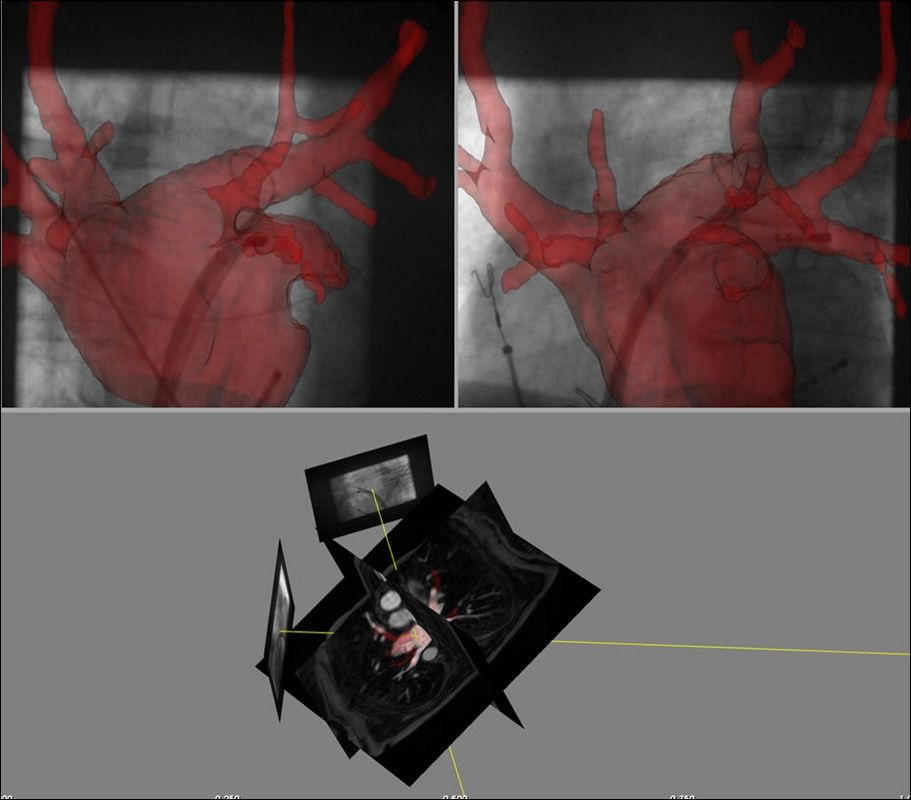
Transvascular procedures are commonly performed under x-ray (XR) fluoroscopy guidance. Providing high temporal and spatial resolution, XR lacks anatomical information. Multimodal intra-procedural three-dimensional (3D) image fusion (IF) can augment the limited information available from XR with relevant anatomic soft-tissue structures. Visualization of anatomical information is achieved by projecting organ shape models derived from e.g. high-fidelity pre-interventional computed tomography angiography (CTA) or magnetic resonance imaging (MRI) data on the live fluoroscopy image.
Objectives:
- To develop a platform for integration of pre-interventional anatomical and functional data with realtime X-ray fluoroscopy data.
- To provide a three-dimensional navigation tool for reproducible 3D positioning of the working catheter and anatomically correct documentation of the working points in 3D during transvascular procedures.
Students:
- Dagmar Bertsche, M.Sc.
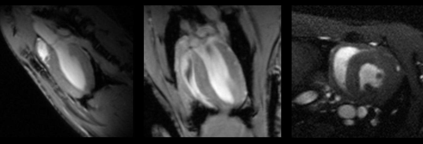
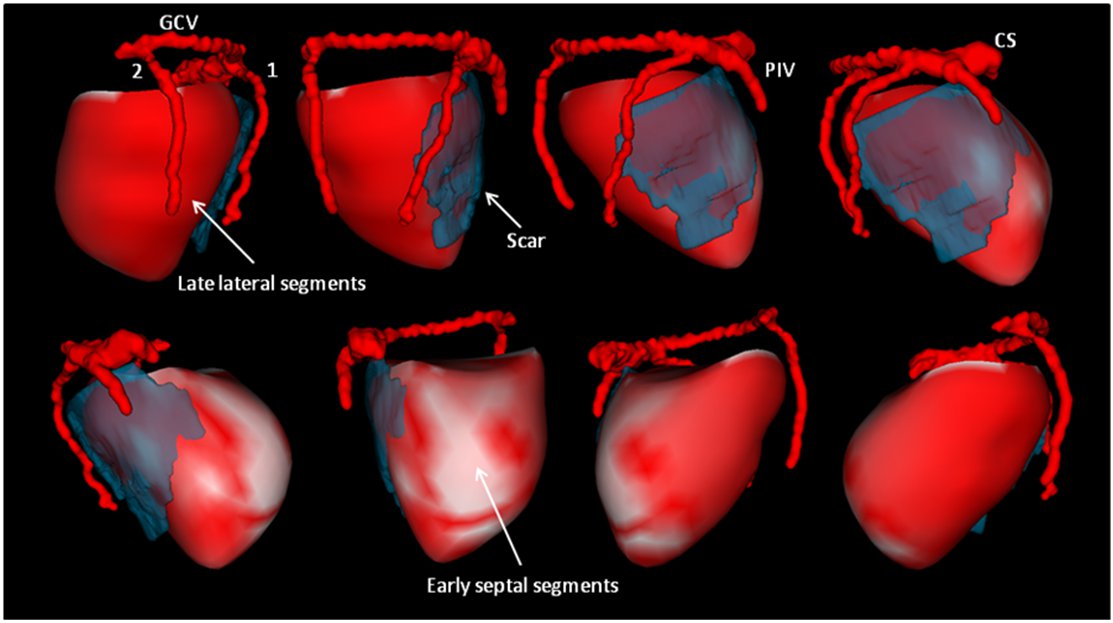
Fusion of anatomic and functional MRI data
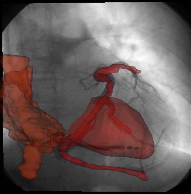
Fusion of pre-interventional data and X-ray fluoroscopy
3D Navigation Tool