Small Animal MRI
Contact: Dr. rer. nat. Alireza Abaei ✉
The Core facility Small Animal Imaging operates an ultrahigh field 11.7T MR imaging and spectroscopy system by Bruker (BioSpec 117/16) equipped with high-sensitive cryogenically cooled receive coils and multi-channel capability. It provides technology for neurological, abdominal and thoracic imaging in mice, rats and tissue samples. The means for physiological gating enables high-quality quantification of cardiac and other functional parameters. Advanced rapid MR spectroscopy (MRS) facilitates the study of a multitude of metabolic processes.
Core Facility Small Animal Imaging
Objectives:
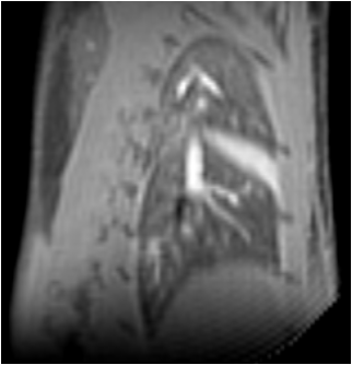
- To investigate high-field functional cardiac imaging
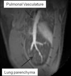
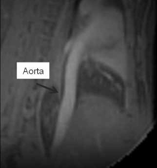
- To investigate high-field ultrashort echo time (UTE) imaging
- To provide support for non-thoracic imaging and spectroscopy
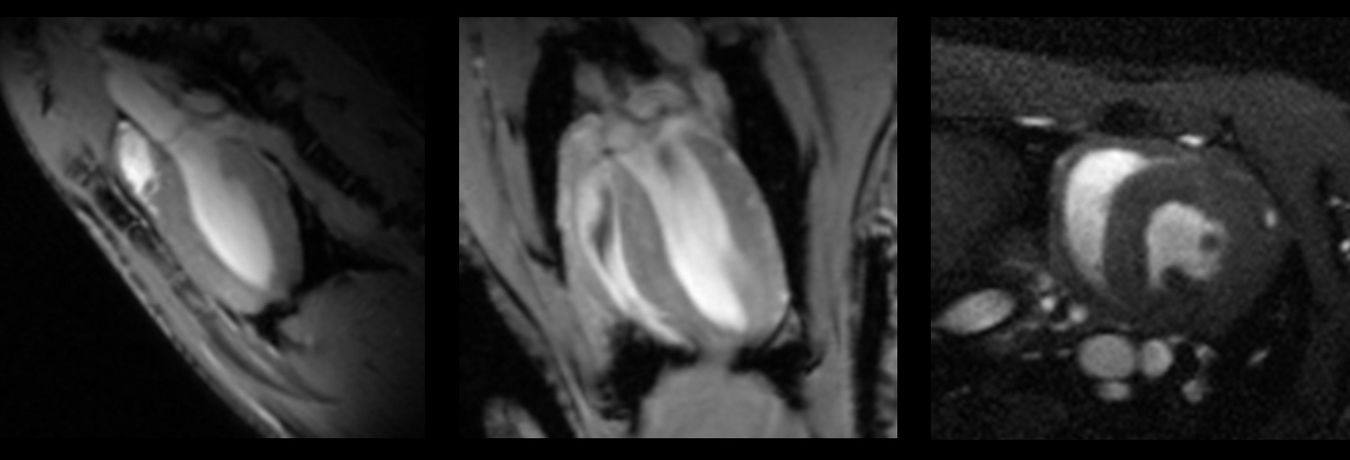
High Resolution Cardiac Imaging
High Resolution Lung Imaging
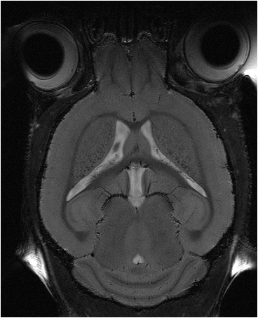
High-Resolution Neuro Anatomy
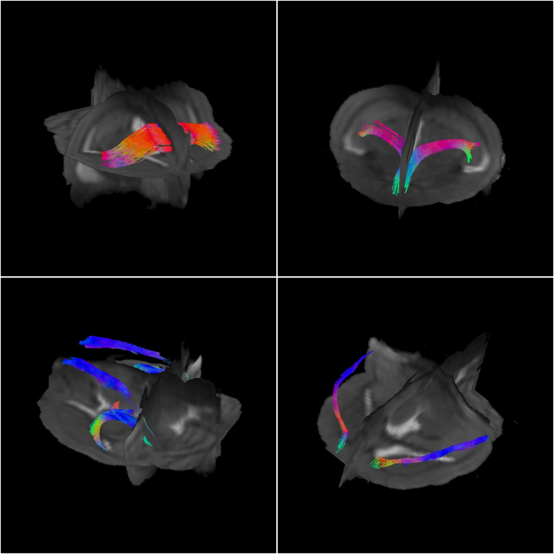
High-Resolution Fast Diffusion Tensor Imaging (DTI)